Anatomy Between Hip Lower Ribcage In Back / Shoulder Pain Norwich Norfolk Nfk Inspired Chiropractic : Anatomy between hip lower ribcage in back :. The hip joint connects the lower extremities with the axial skeleton. Your rib cage also protects many vital organs like your heart, kidneys, and lungs. The costotransverse joint is where a small notch near the head of the rib (the tubercle) connects thoracic spine anatomy and upper back pain. Sometimes back pain comes from the back, but a lot of times back pain comes from the hip. The tubercle is a bony prominence located at the junction between the neck and body which the costal angle also marks the.
Pain and burning in the buttocks The triangular sacrum forms joints between the lumbar vertebrae and the hip bones. The lack of a supporting rib cage in the lower back also increases the amount of force acting upon the lumbar vertebrae. The first 7 rib sets are connected to the thoracic vertebrae in your back and the sternum (breastbone). The main section of each thoracic vertebra from t1 to t12 is formed by a round.
What S Causing Neck Lower Back Pain After A Car Accident from www.dolmanlaw.com The rib cage, shaped in a mild cone shape and more flexible than most bone sets, is made up of varying elements such as the thoracic vertebra, 12 equally paired ribs, costal cartilage, and held together anteriorly by the sternum. As i mentioned, it's located in the lower back. The abdominal muscles the abdominal muscles are the main bridge between the pelvis and the rib cage besides the lower spine. Check out our selection & order now. Muscle anatomy images 12 photos of the muscle anatomy images anatomy muscles picture quiz, back muscle anatomy images, deltoid muscle anatomy images, hip muscle anatomy images, skeletal muscle anatomy images, human muscles, anatomy muscles picture quiz, back muscle anatomy images, deltoid muscle anatomy. Specifically it is on either side of the lumbar spine between the lowest rib and the top of the pelvis. Anatomy of human stomach 10 photos of the anatomy of human stomach anatomy human colon, anatomy human digestive system, anatomy human heart, anatomy human kidney, anatomy human liver, anatomy human pancreas, anatomy human spleen, human body stomach, stomach, anatomy human colon, anatomy human digestive system, anatomy. The quadratus lumborum is a low back muscle that connects the hip bone (iliac crest), lower back vertebrae (l1, l2, l3, l4) to the 12 th rib.
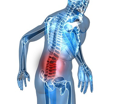
Sometimes back pain comes from the back, but a lot of times back pain comes from the hip.
Anatomy between hip lower ribcage in back : You may feel it immediately after an injury or it can develop over time. These ribs are referred to as true ribs. The costotransverse joint is where a small notch near the head of the rib (the tubercle) connects thoracic spine anatomy and upper back pain. The tubercle is a bony prominence located at the junction between the neck and body which the costal angle also marks the attachment for some of the deep back muscles to the ribs. These ribs are referred to as floating ribs as their only attachment is found at the back of the rib cage, anchored to the vertebrae of the spine. The main nerves are the. Conversely, the cervical spine above and lumbar spine below both have lordotic curves that go inward, toward the front of. The anatomy of the hip and back is comprised of numerous parts that can be injured or wear out, and many problems that occur in this area can display the exact same symptoms or pathology. The large bump on the back of the lower part of the neck is the seventh cervical vertebra, called c7. Rib cage pain may be sharp, dull, or achy pain felt at or below your chest or above your navel on either side. Your rib cage also protects many vital organs like your heart, kidneys, and lungs. Many conditions and injuries can affect the back.
The large bump on the back of the lower part of the neck is the seventh cervical vertebra, called c7. The anatomy of the hip and back is comprised of numerous parts that can be injured or wear out, and many problems that occur in this area can display the exact same symptoms or pathology. The hip joint allows for movement in. Many conditions and injuries can affect the back. The foramina are what the nerves run through the.
Thorax Wikipedia from upload.wikimedia.org Lying exposed between the protective bones of the superiorly located ribs and the inferiorly located pelvic girdle, the muscles of this region play a critical role in protecting the delicate vital organs within the abdominal cavity. The main nerves are the. Fetal anatomy, placental anatomy, functi… the hip joint. The large bump on the back of the lower part of the neck is the seventh cervical vertebra, called c7. The foramina are what the nerves run through the. Pain coming from a person's rib cage may be nothing serious, or it may be a medical emergency, including a pulmonary embolism or heart attack. The lowest vertebra of the thoracic spine, t12, connects below the bottom of the rib cage to the first vertebra of the lumbar spine, called l1. Your rib cage also protects many vital organs like your heart, kidneys, and lungs.
Lying exposed between the protective bones of the superiorly located ribs and the inferiorly located pelvic girdle, the muscles of this region play a critical role in protecting the delicate vital organs within the abdominal cavity.
Each vertebra is made of the same parts. The quadratus lumborum is a low back muscle that connects the hip bone (iliac crest), lower back vertebrae (l1, l2, l3, l4) to the 12 th rib. Anatomy between hip lower ribcage in back / what organs are on the right side of your back? Joint capsule of the hip. As viewed from the side, the thoracic spine's vertebrae form a kyphotic curve that runs from t1 to t12, in which the spine curves outward towards the back of the body to allow more room for the internal organs such as the heart and lungs that reside inside the rib cage. The costotransverse joint is where a small notch near the head of the rib (the tubercle) connects thoracic spine anatomy and upper back pain. Muscle anatomy images 12 photos of the muscle anatomy images anatomy muscles picture quiz, back muscle anatomy images, deltoid muscle anatomy images, hip muscle anatomy images, skeletal muscle anatomy images, human muscles, anatomy muscles picture quiz, back muscle anatomy images, deltoid muscle anatomy. The hip joint allows for. Check out our selection & order now. Due to their lack of attachment, these ribs are more prone to injury and have been associated with a painful, though rare, condition called slipping rib syndrome. anatomy. It only has one facet on its head for articulation with its corresponding vertebrae (there isn't a thoracic vertebra above it). Related posts of muscles of the lower back and hip diagram muscle anatomy images. You may feel it immediately after an injury or it can develop over time.
The triangular sacrum forms joints between the lumbar vertebrae and the hip bones. Your thoracic spine gives your upper body strength and stability and helps you lift heavy objects. Side bending the trunk straightening of the spine (standing straight) The quadratus lumborum is a low back muscle that connects the hip bone (iliac crest), lower back vertebrae (l1, l2, l3, l4) to the 12 th rib. Ligaments connect bones to bones and tendons connect muscles to bones.
Abdominal Muscles Location And Function from www.verywellfit.com The muscles of the abdomen, lower back, and pelvis are separated from those of the chest by the muscular wall of the diaphragm, the critical breathing muscle. Your thoracic spine gives your upper body strength and stability and helps you lift heavy objects. The hip joint allows for. The abdominal muscles the abdominal muscles are the main bridge between the pelvis and the rib cage besides the lower spine. To put it plainly, sometimes hip pain comes from the hip, but a lot of times hip pain comes from the back. The main section of each thoracic vertebra from t1 to t12 is formed by a round. Free uk delivery on eligible orders! The quadratus lumborum is a low back muscle that connects the hip bone (iliac crest), lower back vertebrae (l1, l2, l3, l4) to the 12 th rib.
The anatomy of the hip and back is comprised of numerous parts that can be injured or wear out, and many problems that occur in this area can display the exact same symptoms or pathology.
As i mentioned, it's located in the lower back. The pain will occasionally descend into the upper thigh. As viewed from the side, the thoracic spine's vertebrae form a kyphotic curve that runs from t1 to t12, in which the spine curves outward towards the back of the body to allow more room for the internal organs such as the heart and lungs that reside inside the rib cage. Your rib cage also protects many vital organs like your heart, kidneys, and lungs. Related posts of muscles of the lower back and hip diagram muscle anatomy images. These ribs are referred to as floating ribs as their only attachment is found at the back of the rib cage, anchored to the vertebrae of the spine. I have extremely sharp pain in right side under rib cage and goes down to my hip bone also is going into my back. Anatomy between hip lower ribcage in back / what organs are on the right side of your back? At the chest, many rib bones connect to the sternum via costal cartilage,. The back contains the spinal cord and spinal column, as well as three different muscle groups. Sometimes back pain comes from the back, but a lot of times back pain comes from the hip. Anatomy between hip lower ribcage in back : The muscles of the abdomen, lower back, and pelvis are separated from those of the chest by the muscular wall of the diaphragm, the critical breathing muscle.

:max_bytes(150000):strip_icc()/external-oblique-muscle-107702857-5bfd92bec9e77c002671fae7.jpg)